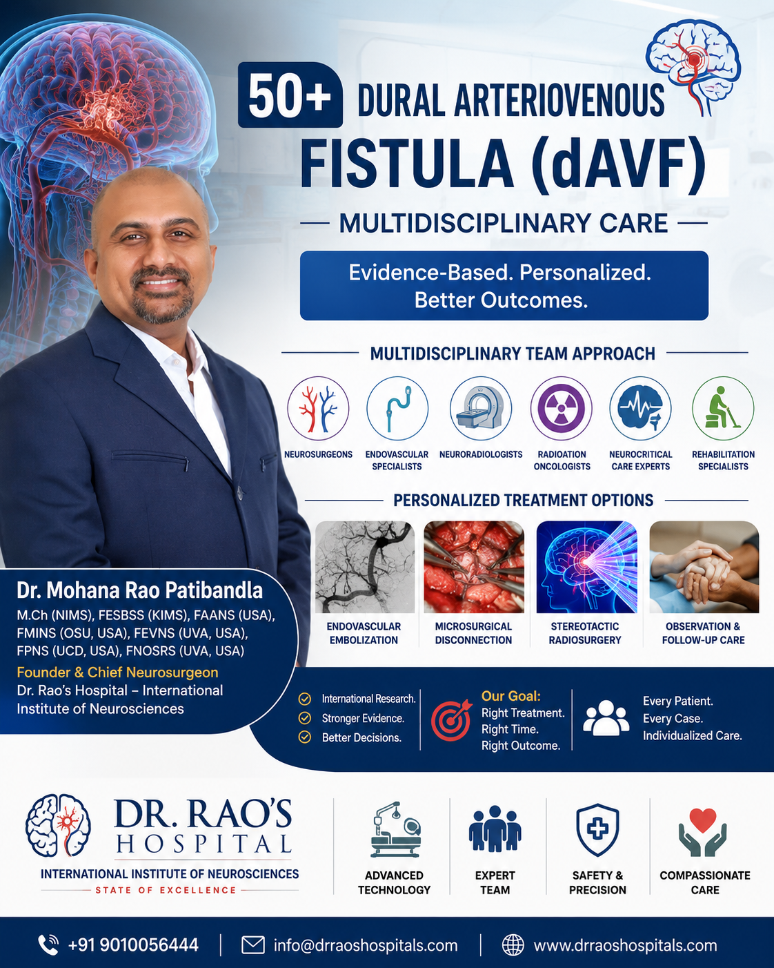
USA-trained neurosurgeon Dr. Rao translates international dAVF research into patient education, explaining personalized care for safer outcomes.
GUNTUR, ANDHRA PRADESH, INDIA, July 29, 2026 /EINPresswire.com/ — A persistent heartbeat-like sound in the ear, unexplained headaches, vision changes, seizures, or even a sudden brain hemorrhage may all have an uncommon but important cause—a dural arteriovenous fistula (dAVF). Although rare, these abnormal connections between arteries and veins surrounding the brain can range from relatively benign conditions to life-threatening vascular disorders. Advances in neuroimaging, endovascular therapy, microsurgery, stereotactic radiosurgery (SRS), and international collaborative research are now enabling physicians to tailor treatment more precisely than ever before.
According to Dr. Mohana Rao Patibandla (Dr. Rao), M.Ch (NIMS), FESBSS (KIMS), FAANS (USA), FMINS (OSU, USA), FEVNS (UVA, USA), FPNS (UCD, USA), FNOSRS (UVA, USA), Founder and Chief Neurosurgeon of Dr. Rao’s Hospital – International Institute of Neurosciences, one of the most significant advances in cerebrovascular care is not simply the availability of new technology, but the ability to select the right treatment for the right patient through evidence-based multidisciplinary decision-making.
Following advanced fellowship training in the United States in cerebrovascular and endovascular neurosurgery, stereotactic radiosurgery, minimally invasive neurosurgery, skull base surgery, pediatric neurosurgery, and neuro-oncology, Dr. Rao contributed to international multicenter research studying the natural history, treatment outcomes, recurrence, and radiosurgical management of dural arteriovenous fistulas. These collaborations through the Consortium for Dural Arteriovenous Fistula Outcomes Research (CONDOR) and the International Gamma Knife Research Foundation continue to improve understanding of this uncommon disease and support more individualized patient care.
Who Is Affected?
Unlike brain arteriovenous malformations, which are usually congenital, dural arteriovenous fistulas are generally acquired vascular disorders that develop later in life. They most commonly affect middle-aged and older adults and may occur after venous sinus thrombosis, trauma, surgery, or other vascular changes, although many patients have no clearly identifiable cause.
Because dAVFs are uncommon and symptoms vary considerably, diagnosis is often delayed. Patients frequently consult ear, eye, or neurology specialists before specialized vascular imaging identifies the underlying condition.
What Is a Dural Arteriovenous Fistula?
A dural arteriovenous fistula is an abnormal connection between arteries and veins located within the dura mater, the protective covering surrounding the brain. Instead of passing through a normal capillary network, blood flows directly into veins under increased pressure.
This abnormal circulation can interfere with normal venous drainage and, depending on the pattern of venous outflow, may lead to progressive neurological symptoms or intracranial hemorrhage.
Importantly, not every dAVF behaves the same way. Some low-risk fistulas may remain stable with observation, while others require prompt intervention because of an increased risk of bleeding or neurological deterioration. International research has demonstrated that the pattern of venous drainage is one of the most important factors in determining both risk and treatment strategy.
When Should Patients Seek Medical Evaluation?
Patients should seek specialist assessment if they experience:
Persistent pulsatile tinnitus (hearing a heartbeat-like sound in the ear)
Unexplained or progressive headaches
Blurred or double vision
Eye redness or swelling
Seizures
Weakness, numbness, or speech difficulty
Problems with balance or coordination
Sudden severe headache or symptoms suggestive of stroke or brain hemorrhage
Early diagnosis allows physicians to evaluate the lesion before irreversible neurological injury occurs and to determine whether observation or treatment is the most appropriate course.
Where and How Should dAVFs Be Managed?
Modern management of dural arteriovenous fistulas requires collaboration between multiple specialists. Treatment decisions may involve endovascular embolization, microsurgery, stereotactic radiosurgery, observation, or a combination of these approaches, depending on the lesion’s anatomy and the patient’s clinical condition.
At Dr. Rao’s Hospital – International Institute of Neurosciences, patients with cerebrovascular disorders are evaluated through a multidisciplinary team that includes cerebrovascular neurosurgeons, endovascular specialists, neuroradiologists, radiation oncologists, neuro-anesthesiologists, neurocritical care physicians, and rehabilitation experts. This collaborative approach reflects contemporary international practice and helps ensure that every patient receives an individualized treatment recommendation rather than a standardized approach.
Dr. Rao’s Philosophy
For Dr. Rao, advances in cerebrovascular medicine are measured not only by technical innovation but by the quality of clinical decision-making.
International research has shown that durable outcomes are achieved when treatment is tailored to the individual patient, guided by scientific evidence, and delivered through multidisciplinary collaboration.
“Every dural arteriovenous fistula presents a different challenge. Our responsibility is to understand the disease, explain the options clearly, and recommend the treatment that offers each patient the safest and most effective long-term outcome. Technology is important, but thoughtful clinical judgment and teamwork remain the foundation of excellent patient care.” — Dr. Mohana Rao Patibandla (Dr. Rao)
International Research Is Shaping Personalized Treatment for Dural Arteriovenous Fistulas
For many years, treatment decisions for dural arteriovenous fistulas (dAVFs) were based largely on anatomical appearance and the experience of individual centers. Because these lesions are relatively uncommon, most hospitals treated only a limited number of patients, making it difficult to answer important questions such as: Which fistulas should be treated? Which treatment is most effective? How often do they recur?
To address these challenges, leading cerebrovascular centers from North America, Europe, and Asia established the Consortium for Dural Arteriovenous Fistula Outcomes Research (CONDOR). By combining data from more than 1,000 patients, the consortium created the world’s largest clinical registry of dAVFs, providing physicians with stronger evidence to guide diagnosis, treatment, and long-term follow-up. Dr. Rao contributed to this international collaborative effort during his fellowship training in the United States, helping translate research findings into practical improvements in patient care.
Endovascular Embolization: Often the First-Line Treatment
Today, endovascular embolization is the preferred initial treatment for many high-grade dAVFs. Through a small catheter inserted into a blood vessel, neurointerventional specialists navigate to the abnormal connection and inject embolic materials to close the fistula while preserving normal circulation.
This minimally invasive technique has transformed cerebrovascular care by reducing the need for open surgery in many patients and allowing faster recovery. However, its success depends on the location and complexity of the fistula.
One of the most important CONDOR studies demonstrated that patients with high-grade, unruptured dAVFs who underwent embolization were more likely to achieve complete obliteration and had a lower risk of hemorrhage and death than similar patients managed conservatively. These findings support embolization as an important first-line option for appropriately selected patients while reinforcing that treatment decisions should always be individualized.
Microsurgery Continues to Play a Vital Role
Although endovascular techniques have advanced significantly, microsurgical disconnection remains an essential part of multidisciplinary cerebrovascular care.
Certain fistulas have complex anatomy that cannot be safely or completely treated through embolization alone. In these situations, surgery offers immediate interruption of the abnormal connection and may provide the most durable cure.
Rather than viewing surgery and embolization as competing approaches, modern practice often combines them. Embolization may reduce blood flow before surgery, making the operation safer, while surgery can definitively treat residual fistulas that remain after endovascular therapy. Selecting the optimal strategy requires careful review of vascular anatomy, venous drainage, neurological symptoms, and overall patient health.
Stereotactic Radiosurgery: A Precise Option for Selected Patients
Not every patient requires open surgery.
Stereotactic radiosurgery (SRS) delivers highly focused radiation to the fistula without an incision. Instead of producing an immediate closure, radiation gradually causes the abnormal vessels to seal over months or years.
Research from the International Gamma Knife Research Foundation, to which Dr. Rao contributed, demonstrated that stereotactic radiosurgery provides durable obliteration with a low risk of permanent complications in appropriately selected patients. SRS is particularly valuable for residual fistulas following embolization or surgery, lesions located in surgically challenging areas, or patients for whom conventional surgery carries increased risk.
Patients should also understand that radiosurgery requires patience. Because the fistula closes gradually, periodic imaging remains essential during the latency period to confirm successful treatment and detect any interval changes.
Observation Versus Intervention
One of the most important messages from international research is that not every dAVF requires immediate treatment.
Low-grade fistulas without cortical venous drainage often have a relatively benign natural history and may be managed safely through careful observation with regular clinical and imaging follow-up. Conversely, high-grade lesions with aggressive venous drainage carry a substantially greater risk of neurological complications or intracranial hemorrhage and usually warrant active treatment.
The decision is therefore not simply whether to operate—it is whether intervention offers greater long-term benefit than observation. Modern treatment planning focuses on balancing procedural risks with the natural history of the individual lesion, ensuring that each patient receives care appropriate to their specific condition rather than a uniform treatment strategy.
Why Long-Term Follow-Up Matters
Many patients believe that treatment ends once imaging shows the fistula has disappeared. However, international collaborative research has demonstrated that recurrence after apparent cure, although uncommon, can occur, particularly in lesions with certain anatomical characteristics.
The CONDOR Consortium identified risk factors associated with recurrence and highlighted the importance of scheduled angiographic follow-up, especially during the first few years after treatment. Early identification of recurrence allows timely retreatment before symptoms or hemorrhage develop.
For patients, this means that successful treatment includes both the initial procedure and continued surveillance as recommended by the treating team.
Why International Collaboration Matters
Rare diseases are difficult for any single institution to study comprehensively. By bringing together leading cerebrovascular centers from around the world, the CONDOR Consortium and the International Gamma Knife Research Foundation have generated evidence that is helping physicians refine treatment strategies, identify risk factors, evaluate long-term outcomes, and improve patient counseling.
For Dr. Rao, participating in these collaborations reinforced an important principle: scientific evidence is most valuable when it improves the care of individual patients.
Rather than applying the same treatment to every dAVF, modern cerebrovascular care emphasizes careful diagnosis, multidisciplinary discussion, and personalized decision-making. Every recommendation should consider the fistula’s anatomy, hemorrhage risk, neurological symptoms, patient preferences, and expected long-term outcome.
“International collaboration has shown us that there is no single solution for every dural arteriovenous fistula. The best outcomes are achieved when scientific evidence, multidisciplinary expertise, and individualized care come together to guide each treatment decision.”
— Dr. Mohana Rao Patibandla (Dr. Rao)
The Future of dAVF Care Is Personalized, Evidence-Based, and Collaborative
The future of dural arteriovenous fistula (dAVF) treatment is being shaped by advances in artificial intelligence (AI), high-resolution vascular imaging, endovascular technology, stereotactic radiosurgery, and precision medicine. These innovations are helping physicians identify complex vascular anatomy more accurately, plan safer procedures, and monitor treatment outcomes with greater confidence.
While technology continues to evolve, Dr. Rao believes that clinical judgment remains at the heart of patient care. Artificial intelligence can assist in analyzing imaging studies and supporting treatment planning, but it cannot replace the experience of a multidisciplinary team or the importance of understanding each patient’s individual circumstances. The best decisions combine scientific evidence, physician expertise, and the patient’s values and goals.
International research has demonstrated that successful management of dAVFs depends on selecting the right treatment for the right patient at the right time. Some lesions require observation, while others benefit from endovascular embolization, microsurgery, stereotactic radiosurgery, or a carefully planned combination of therapies. Long-term follow-up also remains an essential part of care because selected patients may require surveillance imaging even after successful treatment.
“Every dural arteriovenous fistula presents a unique challenge. Our responsibility is to combine scientific evidence, multidisciplinary expertise, and compassionate care to recommend the safest and most effective treatment for each individual patient.”
— Dr. Mohana Rao Patibandla (Dr. Rao)
When Should You Seek Medical Advice?
If you experience persistent pulsatile tinnitus, unexplained headaches, seizures, visual disturbances, neurological deficits, or have been diagnosed with a cerebrovascular vascular malformation, consult a comprehensive neuroscience center for evaluation. Early diagnosis and individualized treatment planning can significantly reduce the risk of complications and improve long-term outcomes.
About Dr. Rao’s Hospital – International Institute of Neurosciences
Dr. Rao’s Hospital – International Institute of Neurosciences, Guntur, is a dedicated neuroscience center providing comprehensive care for disorders of the brain, spine, nerves, and cerebrovascular system. The hospital integrates advanced neuroimaging, cerebrovascular and endovascular neurosurgery, minimally invasive microsurgery, stereotactic radiosurgery, neurocritical care, and rehabilitation, offering evidence-based multidisciplinary care for patients with complex neurological disorders.
Media Contact
Dr. Rao’s Hospital – International Institute of Neurosciences
12-19-67, Old Bank Road, Kothapet, Beside AK Biryani Point, Guntur, Andhra Pradesh – 522001, India
Phone: +91 9010056444
Email: info@drraoshospitals.com | drpatibandla@gmail.com
Website: https://drraoshospitals.com
Selected References
Chen CJ, Buell TJ, Ding D, Patibandla MR, et al. Intervention for Unruptured High-Grade Intracranial Dural Arteriovenous Fistulas. Journal of Neurosurgery. 2022.
Abecassis IJ, Meyer RM, Levitt MR, Patibandla MR, et al. Recurrence After Cure in Cranial Dural Arteriovenous Fistulas (CONDOR). Journal of Neurosurgery. 2022.
Guniganti R, Giordan E, Chen CJ, Patibandla MR, et al. CONDOR Registry: Design and Initial Characterization. Journal of Neurosurgery. 2022.
Starke RM, McCarthy DJ, Chen CJ, Patibandla MR, et al. Evaluation of Stereotactic Radiosurgery for Cerebral dAVFs. Journal of Neurosurgery. 2020.
Starke RM, McCarthy DJ, Chen CJ, Patibandla MR, et al. Hemorrhage Risk Following Gamma Knife Radiosurgery for Cerebral dAVFs. Journal of Neurosurgery. 2020.
Mohana Rao Patibandla
Patibandla Narayana Swamy Neurosciences LLP
+91 90100 56444
email us here
Visit us on social media:
LinkedIn
Instagram
Facebook
YouTube
X
Dr. Rao’s Hosptial – the best neurosurgery hospital in India – with medical tourism
Legal Disclaimer:
EIN Presswire provides this news content “as is” without warranty of any kind. We do not accept any responsibility or liability
for the accuracy, content, images, videos, licenses, completeness, legality, or reliability of the information contained in this
article. If you have any complaints or copyright issues related to this article, kindly contact the author above.

Media gallery